History blog1 July 2026
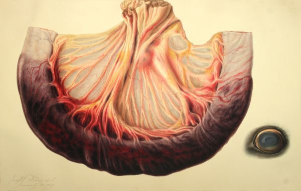
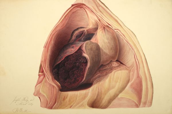
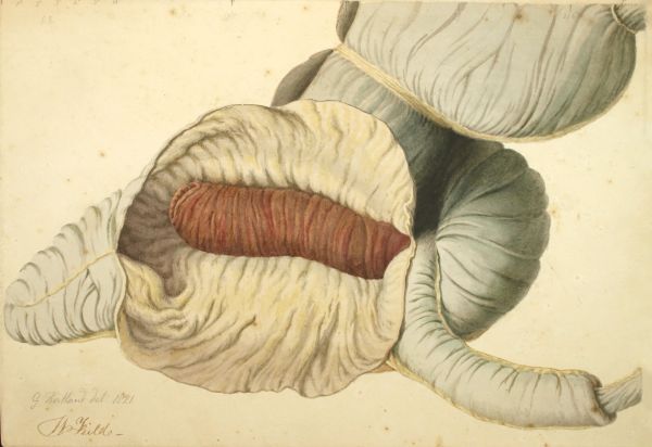
These striking artworks may look like otherworldly landscapes or creatures from the bottom of the sea, but in truth they are illustrations of disease or damage to limbs and organs, mainly in horses. We know that many date from the 1820s and 1830s. They were most likely created to illustrate a publication (as yet unidentified) or as teaching aids.
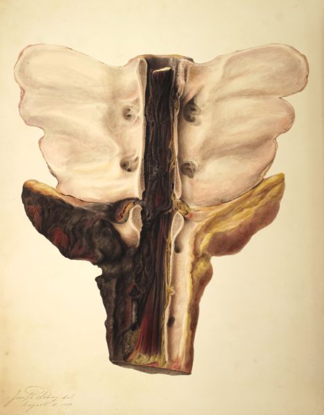
Some are the work of Joseph Perry, an engraver who also created illustrations on human anatomical subjects. Others are signed G Kirtland. One of Kirtland’s trade cards survive in the archives of the British Museum. On it Kirtland describes himself as “Drawing Master and Engraver” of 264 Holborn, London.
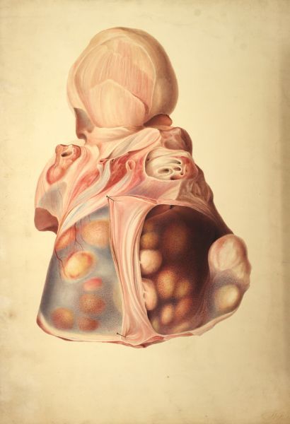
Twenty-one of the paintings are signed by W Field in addition to the name of the artist and it’s assumed these were given to the RCVS by former president William Field (in office 1854-6), as part of a larger donation of items to the Library and Museum made in 1877.
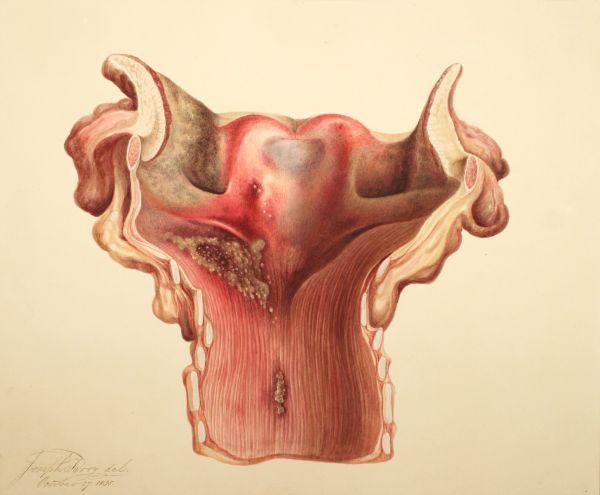
None of this helps us to identify exactly what we’re looking at. Can you assist? Can anybody throw any light as to what these images depict? If so we’d love to hear from you via archives@rcvsknowledge.org
Our puzzles solved
Thank you to those who answered our request for more information about these designs. Thanks to our knowledgable readers we now have a better understanding of these curious images.
Infarction of small intestine. Blackened area is the ischaemic intestine. Blood vessels can be seen within the mesentery. Object bottom right is an eye, so suggesting that in life the mucous membranes were discoloured, leading onto this being a horse.
Three fused lumbar vertebrae [mid-upper of image], itself unusual to that extent, articulated with a sacrum [mid-lower of image]. It is hard to determine what blackened area on the sacrum represents, ?infection? The central cord of dark strands could be a diseased cauda equina. The form of the sacrum suggestive of that of a horse.
This one remains a mystery. Suggestions so far are lumbosacral thrombosis or a possible tapeworm cysts within a serous cavity.
Larynx and trachea, split on ventral aspect, so view is looking dorsally. Two halves of the epiglottis seen as comma-shapes top left and top right. Tracheal rings seen as repeated sections progressing down the image.
Lesion, grey/brown granular, affects right side of laryngeal mucosa plus maybe right half of epiglottis. Hard to say whether the lesion is inflammatory or neoplastic.
Possible haemangiosarcoma on liver or heart. Appears to be within a serous cavity, either to thorax or abdomen. Most likely abdomen, with diaphragm mid-left and greater omentum spilling out at the lower aspect.
Horse caecum, opened to show intussuscepted intestine (brown, ?congested/ischaemic?) within its lumen. The remaining part of the ileum is entering from the bottom right of the image, and can be identified as such by the double mesentery (ileo-caecal ligament plus mesoileum). The entry of the intussusceptum (probably jejunum) into the intussuscipiens (the ileum) can be seen bottom right as the “telescope” effect.
Apex of caecum lower left, base of caecum middle right, right ventral colon top right.
Read more history blogs
-
History blog
 26 August 2026
International Dog Day 2026
-
History blog
-
History blog
 8 June 2026
Open Farm Sunday